Osteoarthritis of the hip, 3D CT scan - stock photo
Coloured computed tomography (CT) scan of the pelvis of a 55 year old patient showing osteoarthritis of the right hip (left). The ball at the top of the femur (thigh bone) fits into the socket of the hip. Osteoarthritis results in the loss of cartilage between the joint. The healing process leads to the growth of bone in place of the cartilage, causing pain, stiffness and loss of mobility. Treatment is with the surgical replacement of the joint

Get this image in a variety of framing options at Photos.com.
PURCHASE A LICENSE
All Royalty-Free licenses include global use rights, comprehensive protection, simple pricing with volume discounts available
$375.00
CAD
Getty ImagesOsteoarthritis Of The Hip 3d Ct Scan High-Res Stock Photo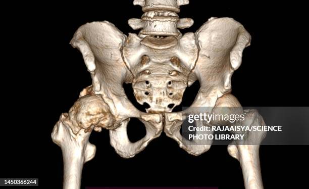 Download premium, authentic Osteoarthritis of the hip, 3D CT scan stock photos from Getty Images. Explore similar high-resolution stock photos in our expansive visual catalogue.Product #:1450366244
Download premium, authentic Osteoarthritis of the hip, 3D CT scan stock photos from Getty Images. Explore similar high-resolution stock photos in our expansive visual catalogue.Product #:1450366244$375$50
Getty Images
In stockDETAILS
Creative #:
1450366244
License type:
Collection:
Science Photo Library
Max file size:
5346 x 3270 px (17.82 x 10.90 in) - 300 dpi - 1 MB
Upload date:
Location:
United Kingdom
Release info:
No release required
Categories:
- Hip - Body Part,
- Joint - Body Part,
- CAT Scan,
- Pelvis,
- Arthritis,
- 3D Scanning,
- Acetabulum,
- Black Background,
- Bone,
- Color Image,
- Color Manipulation,
- Colors,
- Condition,
- Data,
- Degeneration,
- Femur,
- Front View,
- Healthcare And Medicine,
- Horizontal,
- Illness,
- Medical X-ray,
- Medicine,
- No People,
- Osteoarthritis,
- Patient,
- Photography,
- Radiogram - Photographic Image,
- The Human Body,
- Three Dimensional,
- Tomography,
- UK,