antibodies responding to covid-19 coronavirus, illustration - human cell membrane stock illustrationsAntibodies responding to covid-19 coronavirus, illustrationImmune System, Antigen, Virus
cell structure - human cell membrane stock illustrationsCell structureHuman Cell, Mitochondrion, Biology
animal cell, illustration - human cell membrane stock illustrationsAnimal cell, illustrationMitochondrion, Three Dimensional, Nucleus
g protein-coupled receptors, molecular model - human cell membrane stock illustrationsG protein-coupled receptors, molecular modelReceptor, Protein, Dopamine
covid-19 coronavirus binding to human cell, illustration - human cell membrane stock illustrationsCovid-19 coronavirus binding to human cell, illustration
mitochondria, illustration - human cell membrane stock illustrationsMitochondria, illustrationMitochondrion
artwork of mitochondria - human cell membrane stock illustrationsArtwork of mitochondriaMitochondrion, Eukaryotic Cell
destruction of a human cell, illustration - human cell membrane stock illustrationsDestruction of a human cell, illustrationCell Death, Lysis, Human Cell
neuromuscular junction in myasthenia gravis, illustration - human cell membrane stock illustrationsNeuromuscular junction in myasthenia gravis, illustrationAcetylcholine, Receptor, Antibody
chimeric antigen receptor on t-cell, illustration - human cell membrane stock illustrationsChimeric antigen receptor on T-cell, illustrationT-Cell, Receptor, Antibody
mitochondrion, illustration - human cell membrane stock illustrationsMitochondrion, illustrationMitochondrion
ldl particle binding to a receptor, illustration - human cell membrane stock illustrationsLDL particle binding to a receptor, illustrationTriglyceride, Molecule
microscopic view of human b-cells which play a large role in the immune response system. - human cell membrane stock illustrationsMicroscopic view of human B-cells which play a large role in the immune response system.B-Cell
mast cell releasing histamine, illustration - human cell membrane stock illustrationsMast cell releasing histamine, illustrationMast Cell
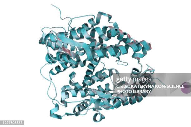
human ace2 receptor molecule - human cell membrane stock illustrationsHuman ACE2 receptor moleculeProtein
covid-19 variant b117, illustration - human cell membrane stock illustrationsCovid-19 variant B117, illustrationCOVID-19, Receptor
human ace2 receptor molecule - human cell membrane stock illustrationsHuman ACE2 receptor moleculeProtein, Molecule, Built Structure
cell structure, illustration - human cell membrane stock illustrationsCell structure, illustrationCilium, Mitochondrion
red blood cells under microscope - human cell membrane stock illustrationsRed blood cells under microscopeHuman Cell, Vector, Microscope
antibodies and covid-19 coronavirus, illustration - human cell membrane stock illustrationsAntibodies and covid-19 coronavirus, illustrationVirus, Antibody
rage receptors in membrane, illustration - human cell membrane stock illustrationsRAGE receptors in membrane, illustrationReceptor, Protein, Diabetes
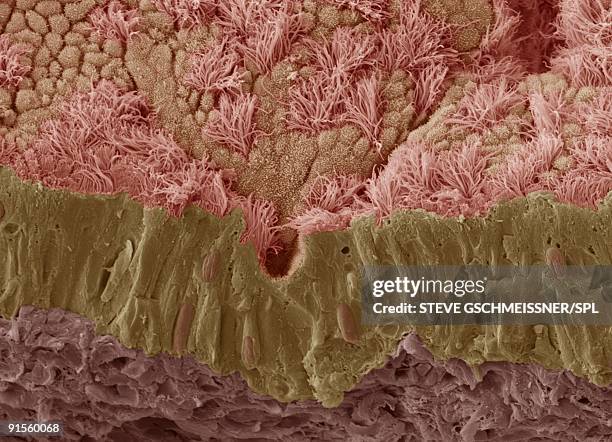
trachea mucous membrane - human cell membrane stock illustrationsTrachea mucous membraneMucus, Human Gland, Illustration
stomach mucosa, light micrograph and illustration - human cell membrane stock illustrationsStomach mucosa, light micrograph and illustrationStomach, Biomedical Illustration
intestinal microvilli, illustration - human cell membrane stock illustrationsIntestinal microvilli, illustrationThree Dimensional
opioid and cannabinoid brain receptors, illustration - human cell membrane stock illustrationsOpioid and cannabinoid brain receptors, illustrationReceptor, Brain, Cell Membrane
insulin binding to insulin receptor - human cell membrane stock illustrationsInsulin binding to insulin receptorMetabolism, Illustration, Molecule
organelles, illustration - human cell membrane stock illustrationsOrganelles, illustrationHuman Cell, Nucleus, Illustration
ldl receptors on cell membrane, illustration - human cell membrane stock illustrationsLDL receptors on cell membrane, illustrationLipoprotein, Triglyceride, High Density Lipoprotein
human cell, illustration - human cell membrane stock illustrationsHuman cell, illustrationHuman Cell, Eukaryotic Cell, Biology
antibodies responding to covid-19 coronavirus, illustration - human cell membrane stock illustrationsAntibodies responding to covid-19 coronavirus, illustrationAntibody, Antigen, Three Dimensional
skin anatomy, illustration - human cell membrane stock illustrationsSkin anatomy, illustrationAnatomy, Built Structure, The Human Body
sars-cov-2 viruses and immune cell, illustration - human cell membrane stock illustrationsSARS-CoV-2 viruses and immune cell, illustration
excitatory synapse, illustration - human cell membrane stock illustrationsExcitatory synapse, illustrationSynapse, Ion Channel, Nerve Cell
antibodies responding to covid-19 coronavirus, illustration - human cell membrane stock illustrationsAntibodies responding to covid-19 coronavirus, illustrationAntibody, Antigen
interleukin 4 binding to its receptor, illustration - human cell membrane stock illustrationsInterleukin 4 binding to its receptor, illustrationReceptor, Cytokine
cell membrane with receptors and ion channels, illustration - human cell membrane stock illustrationsCell membrane with receptors and ion channels, illustrationIon Channel, Opioid
microscopic view of mucus from the nasal mucosa - 19th century - human cell membrane stock illustrationsMicroscopic View of Mucus from the Nasal Mucosa - 19th CenturyEpithelium, Old, Diplococcus
cftr proteins on lung membrane, illustration - human cell membrane stock illustrationsCFTR proteins on lung membrane, illustrationCystic Fibrosis, Epithelium, Healthcare And Medicine
ldl particle binding to a receptor, illustration - human cell membrane stock illustrationsLDL particle binding to a receptor, illustrationMolecule, Triglyceride
mitochondrion, illustration - human cell membrane stock illustrationsMitochondrion, illustrationMitochondrion
cell division - human cell membrane stock illustrationsCell divisionCancer Cell, Three Dimensional, Abstract
anatomy of the organ of corti, part of the cochlea of the inner ear. - human cell membrane stock illustrationsAnatomy of the organ of Corti, part of the cochlea of the inner ear.
microscopic view of human fatty urinary casts from a patient with nephrotic syndrome - 19th century - human cell membrane stock illustrationsMicroscopic View of Human Fatty Urinary Casts from a Patient with Nephrotic Syndrome - 19th Century
covid-19 coronavirus binding to human cell, illustration - human cell membrane stock illustrationsCovid-19 coronavirus binding to human cell, illustration
cell membrane, illustration - human cell membrane stock illustrationsCell membrane, illustrationReceptor, Phospholipid
sars-cov-2 viruses and immune cell, illustration - human cell membrane stock illustrationsSARS-CoV-2 viruses and immune cell, illustrationInfectious Disease, Macrophage, T-Cell
microscopic view of a group of macrophages. macrophages contribute to tumor growth and progression. - human cell membrane stock illustrationsMicroscopic view of a group of macrophages. Macrophages contribute to tumor growth and progression.
microscopic view of plasma cell inside blood vessel. - human cell membrane stock illustrationsMicroscopic view of plasma cell inside blood vessel.Blood Plasma, Pink Color, B-Cell
setmelanotide anti-obesity drug, molecular mode - human cell membrane stock illustrationsSetmelanotide anti-obesity drug, molecular modeBrain, Protein
camp-dependent pathway, molecular model - human cell membrane stock illustrationscAMP-dependent pathway, molecular modelReceptor, Molecule, Dopamine
long term potentiation, illustration - human cell membrane stock illustrationsLong term potentiation, illustration
ion channel in a cell membrane, illustration - human cell membrane stock illustrationsIon channel in a cell membrane, illustration
b cell receptor, illustration - human cell membrane stock illustrationsB cell receptor, illustrationReceptor, Protein, B-Cell
mitochondrion, illustration - human cell membrane stock illustrationsMitochondrion, illustrationMitochondrion, Eukaryotic Cell, Three Dimensional
b.1.617 covid-19 variant, illustration - human cell membrane stock illustrationsB.1.617 Covid-19 variant, illustrationReceptor