covid-19 coronavirus binding to human cell, illustration - human cell membrane stock illustrationsCovid-19 coronavirus binding to human cell, illustration
outer membrane receptor protein molecule - human cell membrane stock illustrationsOuter membrane receptor protein molecule
cardiac sodium channel nav1.5 structure, illustration - human cell membrane stock illustrationsCardiac sodium channel NaV1.5 structure, illustration
sodium-potassium pump in cell membrane, illustration - human cell membrane stock illustrationsSodium-potassium pump in cell membrane, illustration
conceptual image of cell surface receptors. - human cell membrane stock illustrationsConceptual image of cell surface receptors.
microscopic view of human urinary sediment from a patient recovering from hemorrhagic nephritis - 19th century - human cell membrane stock illustrationsMicroscopic View of Human Urinary Sediment from a Patient Recovering from Hemorrhagic Nephritis - 19th Century
melanin, conceptual illustration - human cell membrane stock illustrationsMelanin, conceptual illustration
oxidative phosphorylation, illustration - human cell membrane stock illustrationsOxidative phosphorylation, illustrationAdenosine Triphosphate
nicotinic acetylcholine receptor, illustration - human cell membrane stock illustrationsNicotinic acetylcholine receptor, illustrationReceptor
monkeypox viruses infecting human cell, illustration - human cell membrane stock illustrationsMonkeypox viruses infecting human cell, illustration
cell membrane structure illustration with lipid bilayer and proteins - human cell membrane stock illustrationsCell Membrane Structure Illustration with Lipid Bilayer and Proteins
coxsackievirus bound to human cell, illustration - human cell membrane stock illustrationsCoxsackievirus bound to human cell, illustrationInfectious Disease, Human Cell
potassium-selective ion channel from bacillus cereus, illustration - human cell membrane stock illustrationsPotassium-selective ion channel from Bacillus cereus, illustration
fenestrated capillaries, illustration - human cell membrane stock illustrationsFenestrated capillaries, illustration
structure of mitochondria, illustration - human cell membrane stock illustrationsStructure of mitochondria, illustration
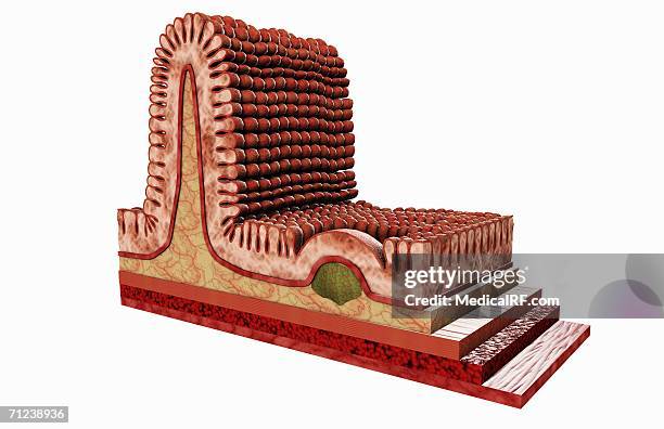
intestine epithelium, illustration - human cell membrane stock illustrationsIntestine epithelium, illustration
mucous cell secreting mucin, illustration - human cell membrane stock illustrationsMucous cell secreting mucin, illustration
b cell receptor dimer, illustration - human cell membrane stock illustrationsB cell receptor dimer, illustrationReceptor
microscopic view of human blood cells and proteus vulgaris bacteria from a patient with peritonitis - 19th century - human cell membrane stock illustrationsMicroscopic View of Human Blood Cells and Proteus Vulgaris Bacteria from a Patient with Peritonitis - 19th Century
microscopic view of human granular urinary casts from a patient with kidney disease - 19th century - human cell membrane stock illustrationsMicroscopic View of Human Granular Urinary Casts from a Patient with Kidney Disease - 19th Century
macrophage engulfing coronaviruses, illustration - human cell membrane stock illustrationsMacrophage engulfing coronaviruses, illustration
her2 and her3 cell membrane receptors forming complex, illustration - human cell membrane stock illustrationsHER2 and HER3 cell membrane receptors forming complex, illustrationProtein
animal cell, illustration - human cell membrane stock illustrationsAnimal cell, illustrationMitochondrion, Illustration, Biology
medical illustration depicting the differences between simple and compound eyes. - human cell membrane stock illustrationsMedical illustration depicting the differences between simple and compound eyes.Compound Eye
amitriptyline antidepressant drug action, illustration - human cell membrane stock illustrationsAmitriptyline antidepressant drug action, illustration
neuromuscular junction in myasthenia gravis, illustration - human cell membrane stock illustrationsNeuromuscular junction in myasthenia gravis, illustrationAutoimmune Disease, Acetylcholine, Receptor
gaba receptors, illustration - human cell membrane stock illustrationsGABA receptors, illustrationBrain, Molecule, Synapse
mitochondria, illustration - human cell membrane stock illustrationsMitochondria, illustrationMitochondrion
antibodies and covid-19 coronavirus, illustration - human cell membrane stock illustrationsAntibodies and covid-19 coronavirus, illustrationAntibody, Immune System
rage receptors in membrane, illustration - human cell membrane stock illustrationsRAGE receptors in membrane, illustrationProtein, Diabetes, Receptor
mast cell, illustration - human cell membrane stock illustrationsMast cell, illustrationMast Cell, White Blood Cell, Autoimmune Disease
insulin binding to insulin receptor - human cell membrane stock illustrationsInsulin binding to insulin receptorInsulin, Illustration, Blood
mast cells, illustration - human cell membrane stock illustrationsMast cells, illustrationIllustration, White Blood Cell, Start Button
interleukin 4 binding to its receptor, illustration - human cell membrane stock illustrationsInterleukin 4 binding to its receptor, illustrationReceptor
camp-dependent pathway, molecular model - human cell membrane stock illustrationscAMP-dependent pathway, molecular modelMolecule, Dopamine, Receptor
covid-19 coronavirus binding to human cell, illustration - human cell membrane stock illustrationsCovid-19 coronavirus binding to human cell, illustration
sars-cov-2 viruses and immune cell, illustration - human cell membrane stock illustrationsSARS-CoV-2 viruses and immune cell, illustration
sars-cov-2 viruses and immune cell, illustration - human cell membrane stock illustrationsSARS-CoV-2 viruses and immune cell, illustration
nmda, ampa and gaba receptors, illustration - human cell membrane stock illustrationsNMDA, AMPA and GABA receptors, illustrationBrain, Ion Channel, Human Nervous System
sars-cov-2 viruses and immune cell, illustration - human cell membrane stock illustrationsSARS-CoV-2 viruses and immune cell, illustration
antibodies and covid-19 coronavirus, illustration - human cell membrane stock illustrationsAntibodies and covid-19 coronavirus, illustration
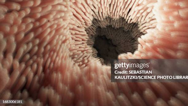
intestinal microvilli, illustration - human cell membrane stock illustrationsIntestinal microvilli, illustration
intestinal microvilli, illustration - human cell membrane stock illustrationsIntestinal microvilli, illustrationThe Human Body
mitochondria, illustration - human cell membrane stock illustrationsMitochondria, illustrationMitochondrion
sars-cov-2 viruses and immune cell, illustration - human cell membrane stock illustrationsSARS-CoV-2 viruses and immune cell, illustration